
Motor neurons, light micrograph (C024/0081) Pin
Neuron Pin
designed and sold by SciencePhoto
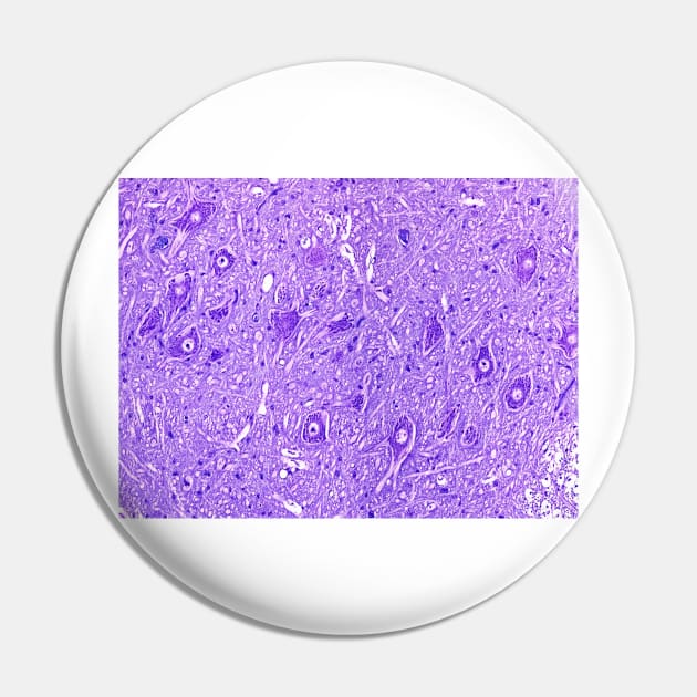
Light microscopy of motor neurons in spinal cord grey matter. The cell bodies of motor neurons show a central nucleus and peripheral cytoplasm (deep purple). In histologic sections processes of dendrites and an axon extend from each cell body. In humans one such axon may be a metre in length if supplying the foot. Tissue between the cell bodies is called the neuropil and is formed of intermingling dendrites, axons, capillaries and glial cells. Magnification x100 when narrow width printed at 10 cm.

More Motor neurons, light micrograph (C024/0081) Products
People Love TeePublic!
Not what you're looking for?
Try another search.
Product Quality
Our Production Team establishes the highest quality standards for third-party printers who participate in the marketplace to ensure that every product sold is perfect.

Neuron Pin FAQ
UPS MI Domestic (6-8 Business Days)
FedEx 2-Day (4-6 Business Days)
Estimates include printing and processing time.More Shipping Info
We want you to love your order! If for any reason you don't, let us know and we’ll make things right.Learn More
Similar to Motor neurons, light micrograph (C024/0081) Pin
More Neuron products
Customers Also Search
More content to explore
Artist's Applied Tags
Trending Tags
The links above have been automatically generated based on tag usage by third-party designers on the TeePublic platform.